Portal Vein Thrombosis: Diagnosis, Anticoagulation, and Treatment Guide
 Aug, 9 2026
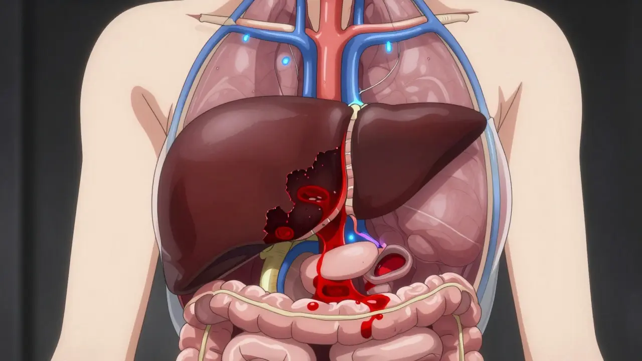
Imagine a traffic jam on the most important highway leading to your liver. That is essentially what Portal Vein Thrombosis is. It happens when a blood clot blocks the portal vein, the large vessel that carries nutrient-rich blood from your intestines to your liver. If left untreated, this blockage can lead to serious complications like intestinal ischemia (tissue death due to lack of blood flow) or worsening portal hypertension. The good news? Early diagnosis and proper anticoagulation therapy can clear the clot in many cases, with survival rates reaching 85% at five years for those treated promptly.
This condition isn't just a rare curiosity; it affects thousands of people annually. Understanding how doctors diagnose it and why they choose specific blood thinners is crucial for patients and caregivers navigating this complex liver health issue. Let’s break down what you need to know about getting diagnosed, choosing the right medication, and managing risks.
Understanding Portal Vein Thrombosis: Acute vs. Chronic
To treat PVT effectively, you first need to understand its timeline. Doctors classify the condition into two main types based on how long the clot has been there:
- Acute PVT: This occurs within the last two weeks. These cases are often painful and come with sudden symptoms like abdominal pain or vomiting. The prognosis is generally better because the clot is fresh and more likely to dissolve with treatment.
- Chronic PVT: This persists beyond six weeks. Over time, the body tries to bypass the blockage by creating new, small blood vessels (collaterals). This process, called cavernous transformation, makes recanalization (reopening the original vein) much harder.
The underlying cause matters too. In non-cirrhotic patients, about 25-30% have an inherited tendency to clot (thrombophilia), such as Factor V Leiden mutation. In cirrhotic patients, the slow blood flow through scarred liver tissue creates a perfect storm for clots to form. Knowing whether you have cirrhosis changes the entire treatment strategy, balancing the risk of bleeding against the risk of the clot growing.
How Is Portal Vein Thrombosis Diagnosed?
You can’t treat what you can’t see. Diagnosis relies heavily on imaging technology. Here is the standard workflow used by hepatologists:
- Doppler Ultrasonography: This is always the first step. It’s non-invasive, cheap, and highly effective. According to recent data, it detects portal vein abnormalities with 89-94% sensitivity. The doctor looks for absent blood flow or reversed flow direction in the portal vein.
- CT or MRI Portography: If the ultrasound is unclear or if the doctor needs to plan surgery, they will order a CT scan with contrast or an MRI. These provide a detailed 3D map of the clot, showing exactly where it starts and ends, and whether it has spread to other veins like the superior mesenteric vein.
- Liver Function Tests: Blood tests check your Child-Pugh score and MELD score. These scores tell the doctor how well your liver is still working despite the clot. A high MELD score indicates severe disease and higher surgical risk.
- Endoscopy: Before starting any blood thinner, especially if you have cirrhosis, you need an endoscopy. This camera test checks for varices (enlarged veins in the esophagus or stomach). If varices are present, they must be banded (tied off) before anticoagulation begins to prevent life-threatening bleeding.
Doctors also classify the severity of the occlusion: minimally occlusive (less than 50% blockage), partially occlusive (50-99%), or completely occlusive (100%). This classification helps predict how well the vein might reopen with treatment.
Anticoagulation Therapy: The Core Treatment
The primary goal of treating PVT is not just to stop the clot from growing, but to encourage the body to dissolve it (recanalization). Anticoagulation, commonly known as blood thinners, is the standard of care for most acute cases. However, choosing the right drug is a delicate balance.
Here are the three main classes of anticoagulants used today:
| Drug Class | Examples | Best For | Key Considerations |
|---|---|---|---|
| LMWH (Low Molecular Weight Heparin) | Enoxaparin (Lovenox) | Initial treatment, Cirrhotic patients (Child-Pugh A/B) | Injected daily. Requires monitoring of anti-Xa levels. Stable effect even with poor liver function. |
| VKA (Vitamin K Antagonists) | Warfarin (Coumadin) | Patients who cannot take DOACs or LMWH | Oral pill. Requires frequent INR blood tests (target 2.0-3.0). Diet affects effectiveness. |
| DOACs (Direct Oral Anticoagulants) | Rivaroxaban (Xarelto), Apixaban (Eliquis) | Non-cirrhotic patients, Selected compensated cirrhosis cases | Oral pill. No routine monitoring needed. Higher recanalization rates in non-cirrhotics. Caution in severe liver disease. |
Why DOACs are gaining popularity: Recent studies, including a 2020 analysis in Blood Advances, show that DOACs like dabigatran and rivaroxaban achieve complete radiographic resolution in 65-75% of non-cirrhotic patients, compared to only 40-50% with older drugs like warfarin. They are easier to take (no injections, no weekly blood tests) and have fewer drug-food interactions.
The Cirrhosis Caveat: If you have cirrhosis, your liver processes drugs differently. Warfarin can be unpredictable because liver disease naturally raises your INR, making it hard to know if you’re over-medicated. LMWH is often preferred initially because its effect is more stable. DOACs are now being used in carefully selected patients with Child-Pugh A or B7 cirrhosis, but they are generally avoided in Child-Pugh C (severe liver failure) due to bleeding risks.

Timing Is Everything: Recanalization Rates
One of the most critical factors in PVT treatment is speed. The longer you wait, the less likely the vein is to reopen. Data from a 2023 study highlights this starkly:
- Within 6 months of diagnosis: 65-75% of patients achieve complete recanalization with anticoagulation.
- After 6 months: Recanalization rates drop to 16-35%. The clot becomes organized and fibrotic, acting more like a solid scar than a soft jelly-like mass.
This is why early detection is vital. If you have risk factors like cirrhosis, inflammatory bowel disease, or a history of cancer, regular screening ultrasounds can catch PVT before it becomes chronic. For transplant candidates, clearing the portal vein is essential. Studies show that anticoagulated transplant candidates have an 85% one-year post-transplant survival rate, compared to 65% for those who were not anticoagulated.
Managing Risks: Bleeding vs. Clotting
The biggest fear for doctors treating PVT is causing a major bleed, particularly from esophageal varices. This is why the pre-treatment checklist is so strict:
- Screen for Varices: Every patient with cirrhosis gets an endoscopy. If medium or large varices are found, they undergo band ligation (tying them off) before starting blood thinners.
- Check Platelet Count: Low platelets (thrombocytopenia) increase bleeding risk. If your count is below 50,000/μL, doctors may transfuse platelets temporarily to safely start LMWH.
- Assess Ascites: Uncontrolled fluid buildup in the abdomen (ascites) suggests advanced liver decompensation. Anticoagulation is usually contraindicated here unless the ascites is managed.
Even with precautions, major bleeding occurs in 5-12% of cirrhotic patients on anticoagulation, mostly from variceal sources. In non-cirrhotic patients, this risk drops to 2-5%. If bleeding does occur, reversal agents like andexanet alfa (approved in 2023) can quickly neutralize certain DOACs, providing a safety net that didn’t exist a few years ago.

When Medication Isn’t Enough: Advanced Interventions
If anticoagulation fails, or if the clot is causing immediate threats like bowel ischemia, doctors may turn to invasive procedures:
- TIPS (Transjugular Intrahepatic Portosystemic Shunt): A radiologist places a stent inside the liver to create a shortcut between the portal vein and hepatic vein. This lowers pressure and can help clear the clot. Success rates are 70-80%, but it carries a 15-25% risk of hepatic encephalopathy (confusion due to toxins reaching the brain).
- Mechanical Thrombectomy: Using catheters to physically suck out or break up the clot. This is reserved for specialized centers and offers 60-75% immediate recanalization, but it’s technically demanding.
- Surgical Shunts: Rarely used today due to high complication rates, but considered if TIPS fails.
These interventions are typically second-line options. The first line is almost always anticoagulation, provided the patient isn’t actively bleeding.
Duration of Treatment: How Long Do You Stay on Thin Ice?
There is no one-size-fits-all answer here. The duration depends entirely on why the clot formed:
- Provoked PVT (Temporary Risk Factor): If the clot was caused by a temporary issue like surgery, infection, or pregnancy, treatment lasts for a minimum of 6 months. Once the risk factor is gone and the vein is open, you can stop.
- Unprovoked PVT (Thrombophilia): If you have an inherited clotting disorder (like Factor V Leiden) or active cancer, lifelong anticoagulation is usually recommended. Stopping could lead to recurrence.
- Cirrhotic PVT: Guidelines are evolving. Many experts recommend continuing until the vein recanalizes or indefinitely if the patient is awaiting a transplant. Recent updates suggest considering discontinuation after 6-12 months if the clot resolves and liver function stabilizes, but this requires close monitoring.
Regular follow-up ultrasounds every 3-6 months are standard to monitor for recurrence or progression. Don’t stop your medication without discussing it with your hepatologist.
What are the first signs of portal vein thrombosis?
Acute PVT often presents with sudden, severe abdominal pain, nausea, vomiting, and sometimes bloody stools if intestinal ischemia is occurring. Chronic PVT may be asymptomatic or present with signs of portal hypertension, such as ascites (fluid in the belly), splenomegaly (enlarged spleen), or variceal bleeding. Many cases are discovered incidentally during ultrasounds for other conditions.
Can portal vein thrombosis be cured?
Yes, in many cases. "Cure" in this context means recanalization-the reopening of the blocked vein. With early anticoagulation, 65-75% of patients achieve complete recanalization. Even if the vein doesn’t fully reopen, controlling the clot prevents life-threatening complications like bowel necrosis or liver failure.
Is it safe to take blood thinners if I have cirrhosis?
It can be, but it requires careful management. Patients with Child-Pugh A or B cirrhosis often benefit from anticoagulation, which improves transplant outcomes. However, they must undergo endoscopic screening and banding of varices first to reduce bleeding risk. LMWH is often preferred initially, while DOACs are used selectively. Child-Pugh C patients are generally excluded due to high bleeding risks.
Which blood thinner is best for PVT: Warfarin or DOACs?
For non-cirrhotic patients, DOACs (like rivaroxaban or apixaban) are increasingly preferred due to higher recanalization rates and ease of use. For cirrhotic patients, LMWH (enoxaparin) is often the safest initial choice due to predictable pharmacokinetics. Warfarin is less favored now due to the need for frequent monitoring and dietary restrictions, though it remains an option for those who cannot afford or tolerate newer drugs.
How long do I need to stay on anticoagulation?
The duration depends on the cause. If triggered by a temporary event (like surgery), 6 months is typical. If caused by an inherited clotting disorder or active cancer, lifelong therapy is usually recommended. For cirrhotic patients, treatment continues until recanalization occurs or indefinitely if awaiting transplant, guided by individual risk assessments.

Dennis Leeftink
August 10, 2026 AT 03:58Most people here dont get it. The liver is not just a filter, it is the main chemical plant of the body. If you block the portal vein you are stopping the fuel supply. Warfarin is old school garbage for this specific issue because the liver makes the clotting factors and if the liver is sick the INR lies to you. You need LMWH or DOACs if you can afford them. Simple as that.
Ambria St louis
August 11, 2026 AT 15:59Oh my goodness! It really is fascinating how the body tries to compensate by creating those collaterals! I mean, think about the sheer engineering marvel of cavernous transformation! It is quite literally the body trying to bypass a roadblock! And yet, we often overlook the importance of early screening! We should all be more vigilant about our liver health!
Michelle Alavaski
August 12, 2026 AT 00:59One must consider the possibility that the pharmaceutical industry has a vested interest in promoting anticoagulants over natural blood thinners such as nattokinase or high-dose vitamin C. The data presented here relies heavily on studies funded by drug manufacturers who stand to profit from long-term prescriptions. It is highly probable that the 'standard of care' is designed to keep patients dependent rather than cured. Skepticism is warranted when survival rates are cited without disclosing the quality of life metrics for those surviving five years.
Veronica Agbanyim
August 12, 2026 AT 21:54It is morally imperative that healthcare systems prioritize early detection for at-risk populations. Too many people suffer in silence because they lack access to basic ultrasound screening. The disparity in outcomes between those who receive prompt treatment and those who do not is a reflection of systemic neglect. We have a duty to ensure that financial status does not determine whether one receives life-saving anticoagulation therapy. Justice demands equitable access to these diagnostics.
John Divers
August 13, 2026 AT 16:10The distinction between acute and chronic presentation is vital for clinical decision-making. Early intervention yields significantly better recanalization rates. Collaboration between hepatologists and radiologists ensures optimal imaging protocols are utilized promptly.
jackie healey
August 14, 2026 AT 05:16As a nurse who has worked with several PVT patients! I can tell you that the fear of bleeding is very real for everyone involved! But knowing that banding varices first reduces that risk so much gives me peace of mind! It is wonderful to see guidelines evolving to include DOACs for non-cirrhotic patients! It makes their lives so much easier without daily injections!
Traci Bobbitt
August 15, 2026 AT 19:38Look, nobody likes getting poked. If you are non-cirrhotic and your doctor offers Eliquis or Xarelto, take it. Warfarin is a pain in the butt because you have to watch every green vegetable you eat. Just make sure you check your platelets though. Low platelets mean higher bleed risk. That is the only catch. Otherwise, the new drugs work way better for clearing the clot.
Sansaray Jones
August 17, 2026 AT 13:07interesting read. the timeline matters most. if you wait too long the clot hardens. simple fact.
Anna Salamon
August 18, 2026 AT 16:11In many cultures, abdominal pain is dismissed as indigestion until it becomes severe. This delay can be catastrophic for conditions like Portal Vein Thrombosis. Education is key. Patients must understand that sudden, severe abdominal pain requires immediate medical attention, not home remedies. Supporting patient advocacy groups helps spread this critical awareness globally.